
Hiranonis and Prevotella copri have strong negative relationships between abundance and species richness b 02. ConspectusThe last decades have witnessed unprecedented scientific breakthroughs in all the fields of knowledge from basic sciences to translational research resulting in the drastic improvement of the lifespan and overall quality of life.
Solved Calculate The Concentration Of Proteins In The Final Chegg Com
Standard Curve Wikipedia
B 7 Analysis Of Protein Concentration Hl Youtube
The increase of absorbance at 595 nm is proportional to the amount of bound dye and thus to the amount concentration of protein present in the sample.
Relationship between concentration and absorbance of protein in spectrometry. See also spectral linewidth. The sequence of amino acids determines each proteins unique 3-dimensional structure and its specific function such as catalysis of biochemical reactions mechanical support and immune protection. NMR measurements of p107 612-687 were recorded using either 15 N- or 15 N 13 C-labeled protein at a final concentration of 6 or 200 μM in NMR buffer 20 mM sodium phosphate pH 68 250 or 50 mM NaCl 05 mM TCEP and 90 H 2 O10 D 2 O.
When sample absorbance exceeds about 005 in a 1 cm pathlength the relationship becomes nonlinear and measurements may be distorted by artifacts such as self-absorption and the inner-filter effect. We would like to show you a description here but the site wont allow us. Schaap et al 2002.
There is a linear relationship between the absorbance and the amount of sugar that was present in the original sample. Because fluorescence quantitation is dependent on the instrument fluorescent reference standards are essential for calibrating measurements made at different times or using different instrument. However despite these great advances the treatment and diagnosis of some diseases remain a challenge.
However lipids in patient specimens are far more complex than in Intralipid hence there is a poor relationship between triglyceride concentration and LI in patient samples. The aim of the present study was to establish a possible relationship between biofilm-forming capacity and the antibiotic-resistant phenotype in. The decay constant of the experimental data fits reveals that C.
This method determines both reducing and non-reducing sugars because of the presence of the strongly oxidizing sulfuric acid. Unlabeled B55α and 15 N-labeled p107 was mixed at 11 ratio to form the complex. Left-hand circular LHC and right-hand circular RHC polarized light represent two possible spin angular momentum states for a photon and so circular dichroism is also referred to as dichroism for spin angular momentum.
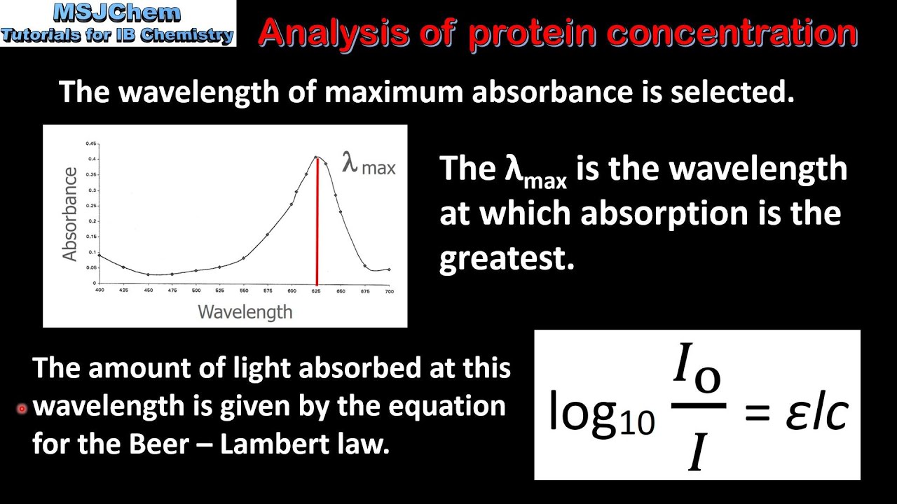
Antibiotic resistance in Staphylococcus aureus is associated with reduced neutrophil recruitment. A plot of the response as a function of wavelengthor more commonly frequencyis referred to as a spectrum. Spectrometry is the spectroscopic technique used to.
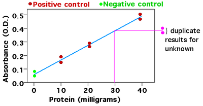
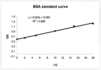
Any absorbance observed in this process is to be subtracted from the absorbance of the sample. Xy-0067710736 so x0495mgml As you can see by the Linear plot number above a linear regression does not provide a good method for comparing the protein standards to the unknown samples. Inspired by nature scientists have.
The protein belongs to the fatty-acid-binding protein the prevalent childhood diseases of jaundice and kernicterus. Here Payne et al. Combined with the application of the above screening technologies in S protein-ACE2 blockers a variety of natural products such as flavonoids terpenoids phenols alkaloids were summarized which could be used as S protein-ACE2 blockers in order to provide ideas for the efficient discovery of S protein-ACE2 blockers from natural sources and contribute to the development of broad-spectrum.
The absorbance of the sample is noted in different frequencies which usually ranges from 200-800 nm. Then cells were switched to an unlabeled medium supplemented with 50 gL albumin either with or without leucine and after the indicated amount of time cellular protein was extracted and analyzed by quantitative mass spectrometry which differentiates between old labeled protein and new unlabeled protein Figure 5A. Tyrosol is a phenolic found in extra virgin olive oil EVOO.
Citation needed Unlike other protein assays the Bradford protein assay is less susceptible to interference by various chemical compounds such as sodium potassium or even carbohydrates like sucrose that may be present in protein samples. Solving for x the protein concentration for an absorbance of 06 gives. The relationship between the multidrug-resistant MDR phenotype and biofilm-forming capacity has been a topic of extensive interest among biomedical scientists as these two factors may have significant influence on the outcomes of infections.
Concentration dependence of the absorbance at the oscillator position 1700 cm 1 black curve compared to the normalized absorbance green line integrated from 1003000 cm 1 to the normalized integrated absorbance calculated between 16001800 cm 1 red line and to the normalized integrated absorbance calculated between 16001840 cm 1 blue line. With enzymes or 2 volume displacement by lipid causing a decreased aqueous phase eg. Circular dichroism CD is dichroism involving circularly polarized light ie the differential absorption of left- and right-handed light.
To quantify the relationship between abundance and richness for each species we fit an exponential decay model y ae bx to the data solid colored lines and simulations dashed gray lines. FABP family Storch and Thumser 2000. The HL-60 acute myeloid leukaemia cell line which can be differentiated to both monocytes and neutrophils was exposed to tyrosol at this concentration and analysed for evidence of differentiation and effects of cytotoxicity.
However it is a convenient method providing a rough estimation. The cuvette with the sample solution is then placed in the spectrometer. In a Maltese monocultivar EVOO it was present at a concentration of 923 ppm.
Particle size can be determined by measuring the random changes in the intensity of light scattered from a suspension or solution. A similar spectrum is formed from a different concentration of the samples. The solution is then allowed to cool and its absorbance is measured at 620 nm.
An aliquot of the light phase is removed and its concentration determined spectroscopically eg by measuring absorbance at 280 nm in which case the solution should be diluted to prevent reformation of droplets or with a colorimetric assay or fluorescence intensity. Link formylated peptides which act as. Relationship E hν for photons was realized h is the Planck constant.
In general the technique is best used for submicron particles and can be used to measure particle with sizes less than a nanometer. This technique is commonly known as dynamic light scattering DLS. We determined the structure of holoUnaG at 12 Å and identified Zimmerman and Veerkamp 2002 with the highest homology the bilirubin-binding site as a biplanar p-conjugated structure 56 to brain B- FABP Figure 1A.
The interference mechanism is due either to 1 light scatter causing measurement errors in photometric methods eg.
Exam2 Spectrophotometry Key

Mass Spectrometric Conjugate Characterization Process Qualification Of Recombinant Protein Hapten Conjugation Bioprocess Internationalbioprocess International
1

Protein Quantification Unchained Labs
Chapter 3 Investigating Proteins Chemistry

Absorbance Measurements Bmg Labtech
Molecular Endocrinology Laboratory Spectrophotometry

Molecules Free Full Text Evaluation Of Peptide Protein Self Assembly And Aggregation By Spectroscopic Methods Html


